 info@labtron.com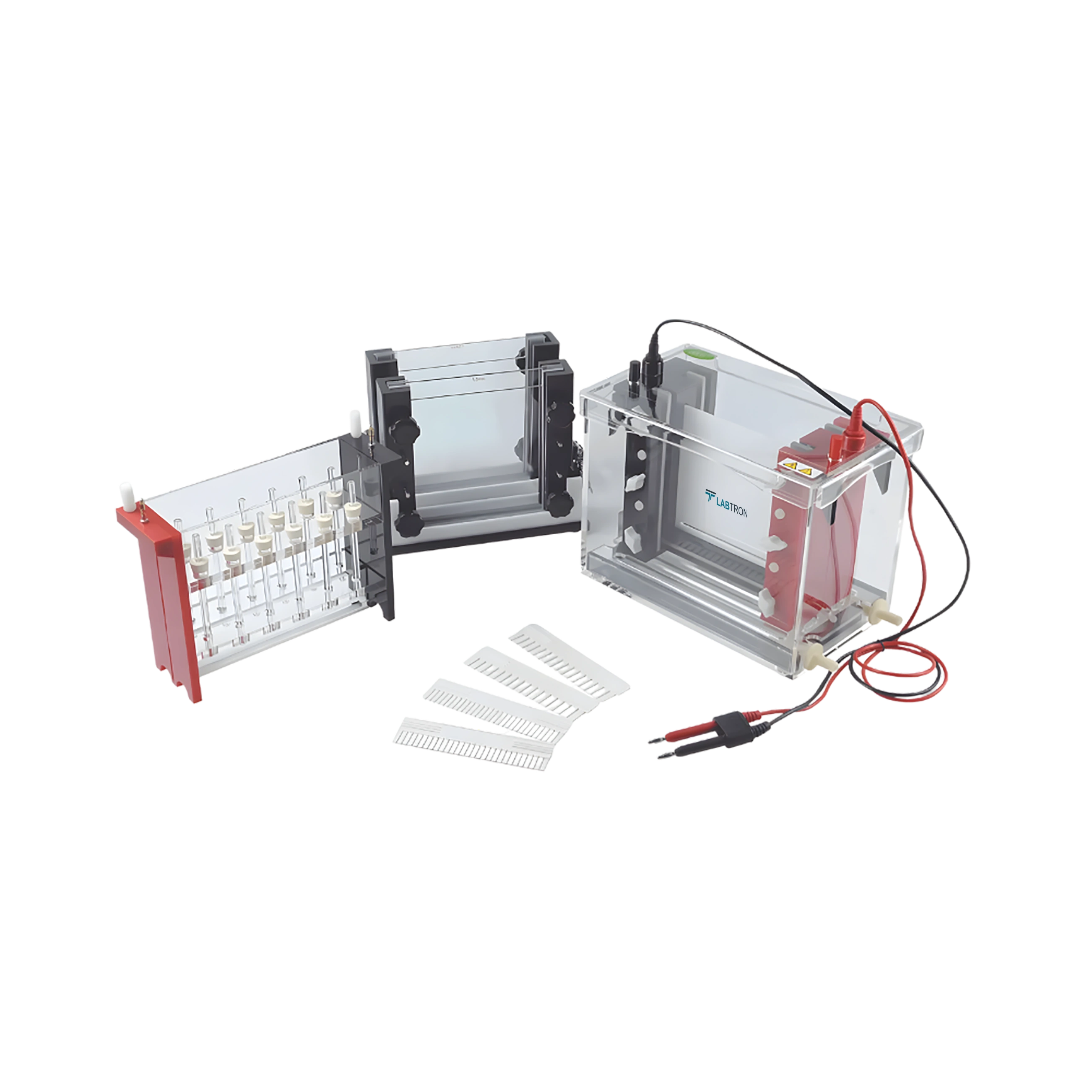

| Length of 1st D Tube gel Electrophoresis | 120 mm |
| Gel Diameter of 1st D Tube Gel Electrophoresis | 1.0 mm, 2.00 mm |
| Gel Dimensions of 2nd Vertical Electrophoresis (W × L) | 143 × 139 mm |
| Gel Thickness of 2nd Vertical Electrophoresis | 1.0 mm, 2.00 mm |
| Number of Gels | 2 pcs |
| Maximum Buffer Volume | 2500 ml |
| Sample Throughput | 17 to 58 |
| Dimension (L × W × H) | 460 × 230 × 400 mm |
| Weight | 13.2 kg |
2D Gel Electrophoresis System LDGES-A10 is a specialized technique to separate complex protein mixtures based on molecular weight and isoelectric point. It is widely used in proteomics research, and functional genomics to analyze protein expression and identify biomarkers.
Download catalog for
2D Gel Electrophoresis System LDGES-A10 Catalog
In 2D Gel Electrophoresis, gel preparation involves two main steps: preparing the isoelectric focusing (IEF) gel for the first dimension and the SDS-PAGE gel for the second dimension. The IEF gel is used to separate proteins by their isoelectric point, and it is typically prepared using polyacrylamide gels with a pH gradient. After the first dimension, the IEF gel is laid on top of the SDS-PAGE gel, and the proteins are separated by their molecular weight in the second dimension. Labtron’s 2D Gel Electrophoresis Systems are designed to simplify and optimize the gel preparation process, ensuring reproducible results and efficient protein separation.
The primary benefit of 2D Gel Electrophoresis over one-dimensional methods is the higher resolution and the ability to separate proteins based on two different parameters (isoelectric point and molecular weight). This allows for the separation of thousands of proteins in a single experiment, offering significantly better proteome coverage. Additionally, it enables the analysis of complex biological samples where many proteins are present in similar molecular weight ranges. Labtron’s 2D Gel Electrophoresis Systems provide exceptional resolution and sensitivity, ensuring researchers can analyze complex protein mixtures with precision.
The 2D Gel Electrophoresis process typically takes several hours to a full day, depending on the complexity of the sample and the specific protocols being used. The first dimension (isoelectric focusing) can take several hours, depending on the gradient used and the type of proteins being separated. The second dimension (SDS-PAGE) usually takes a few hours. After running the gels, additional steps such as staining and imaging can also take additional time. Labtron’s 2D Gel Electrophoresis Systems are designed for efficient, high-throughput workflows, allowing for rapid processing while maintaining high-quality results.
Yes, 2D Gel Electrophoresis can be used for quantitative protein analysis. After protein separation, the intensity of the spots or bands on the gel can be quantified using imaging systems that measure the light absorption or fluorescence of stained proteins. This allows for the comparison of protein expression levels across different conditions or samples. Labtron’s systems are equipped with advanced image analysis software that can perform spot detection, quantification, and normalization, providing accurate and reproducible quantitative data from 2D gels.
Several types of stains can be used in 2D Gel Electrophoresis, including Coomassie Brilliant Blue, silver stain, and fluorescent dyes. Coomassie Brilliant Blue is commonly used for general protein detection due to its sensitivity and ease of use. Silver stain is more sensitive and can detect proteins at lower concentrations, but it is more labor-intensive. Fluorescent dyes, such as SYPRO Ruby, offer even higher sensitivity and can be used for multiplexing in mass spectrometry applications. Labtron’s 2D Gel Electrophoresis Systems are compatible with all these staining techniques, providing flexibility based on the researcher’s needs.
The first dimension, Isoelectric Focusing (IEF), is crucial because it separates proteins based on their isoelectric point (pI). Each protein has a unique pI at which it is electrically neutral, and IEF takes advantage of this by applying a pH gradient to the gel. As the sample is subjected to an electric field, proteins migrate until they reach the point where their net charge is zero (their pI). This step allows for highly efficient separation of proteins, especially when dealing with complex samples. Labtron’s systems optimize IEF by providing precise control over the pH gradient and electrical field, ensuring high-resolution protein separation.
Post your question?
Our experts are here to help!
Labtron Equipment Ltd., Quatro House, Lyon Way, Camberley, Surrey GU16 7ER United Kingdom
Email: info@labtron.com Whatsapp: +44 73 4441 2688 Phone: +44 2080 043608