 info@labtron.com

| Scanning Capacity | 1 slide |
| Slide Size | Supports 26 × 76.5 mm slides |
| Resolution | 20x: up to 0.25 μm/pixel; 40x: up to 0.17 μm/pixel |
| Scanning Time | Up to 45 seconds per slide |
| Operation Mode | Continuous operation |
| Scanning Type | Brightfield |
| Scan Method | Area scanning with autofocus |
| Scan Mode | Single-layer, Z-stack, Extended focus |
| Layer Capacity | Up to 41 layers |
| Scan Area | Automatic identification / Manual selection |
| Objective | PLAN-APOCHROMAT 20x / 40x |
| 100X Oil Support | Not supported |
| Camera | Basler 5 MP CMOS, 75 FPS |
| Light Source | LED |
| Barcode Recognition | 1D and 2D barcode |
| Digital Slide Format | ZYP, JPEG, PNG, BMP, TIFF, SVS, DICO |
| Software Version | V1.1 |
| Electric Shock Protection | Class I |
| Pollution Class | Class II |
| Power Supply | 100 to 240 VAC, 50 to 60 Hz |
| Power Consumption | 80 VA |
| Dimension | 400 × 500 × 600 mm |
| Weight | 35 kg |
Feature | Description |
Scanner Control Software | 1 PC |
Scanner Viewing Software | 1 PC |
Workstation (DEII) Specifications | 20-core Intel Core i7 CPU, 16GB RAM |
Storage | 512GB SSD + 2TB HDD |
Operating System & Media Support | Windows 11 64-bit, DVD-RW drive included |
Our Digital Pathology Slide Scanner LDPS-A17 is designed for high-resolution scanning in clinical and research labs. It supports routine slide digitization, remote diagnostics, and frozen section evaluation in compact lab environments.
Accessory Name | Quantity |
Power Adaptor | 1 pc |
Dongle | 1 pc |
Download catalog for
Digital Pathology Slide Scanner LDPS-A17 Catalog
Download manual for
Digital Pathology Slide Scanner LDPS-A17 Manual
Available Range :
Digital Pathology Slide Scanner LDPS-A10 : 1 Digital Pathology Slide Scanner LDPS-A17 : 1 slide Digital Pathology Slide Scanner LDPS-A11 : 12 Digital Pathology Slide Scanner LDPS-A16 : 120 Digital Pathology Slide Scanner LDPS-A13 : 20 Digital Pathology Slide Scanner LDPS-A14 : 40 Digital Pathology Slide Scanner LDPS-A15 : 60Labtron offers a range of digital pathology slide scanners designed to digitize glass slides containing tissue specimens. They have high-resolution CMOS sensors that achieve rapid scanning times. The dynamic pre-focusing system adjusts the lens in real time, optimizing focus points for clear slide images. Their Z-Stack extended focus captures 3D digitized images of specimens like cell clumps and thick tissues. Our digital pathology slide scanners automatically recognize different slide types, making them ideal for histopathology and more.


.webp "Digital Pathology Slide Scanner LDPS-A10")

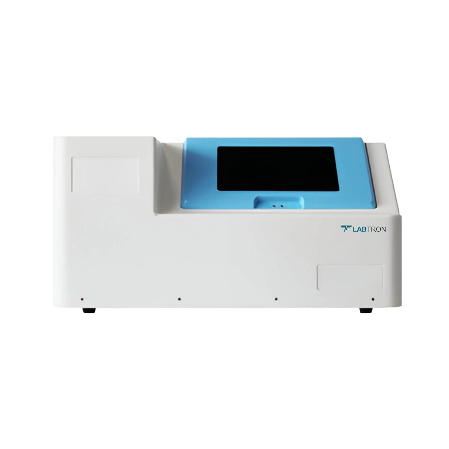


Labtron’s Digital Pathology Slide Scanner uses a Plan-Apochromat objective and a high-speed CMOS camera to deliver sharp, detailed images. Enhanced image algorithms and LED illumination boost contrast and reduce noise. Autofocus and Z-stack functions allow accurate imaging across tissue layers. These enhancements contribute to reliable and consistent image output.
The Digital Pathology Slide Scanner supports multi-window comparison for up to 9 slides at once. This is especially useful for teaching, diagnostic reviews, and cross-case comparisons. Each slide can be annotated and adjusted independently. The interface allows users to toggle between magnifications and color settings per window.
The Digital Pathology Slide Scanner supports single-layer scanning, Z-stack, and extended focus modes. Z-stack captures multiple image layers, which can be reviewed or merged. Extended focus mode combines the clearest parts from each layer into a single image. These options improve clarity and depth for various sample types.
Labtron’s Digital Pathology Slide Scanner provides image processing features such as impurity removal, hole filling, and boundary expansion. These tools improve scan clarity and reduce false detections. Settings can be customized based on sample characteristics. The software ensures optimized results even in complex tissue samples.
The outer surface of the Digital Pathology Slide Scanner should be cleaned with a dry or lightly alcohol-dampened cloth. Liquids should never be sprayed directly onto the device. The slide holder and optical components must be kept free of debris. Internal maintenance should only be conducted by qualified service personnel.
The Digital Pathology Slide Scanner operates at ambient temperatures of 5 °C to 40 °C and humidity upto 80%. Power requirements are 100 to 240 VAC, 50/60 Hz, with 80 VA consumption. A stable, grounded power connection is necessary. Proper environmental conditions ensure consistent scanner performance and equipment longevity.
The Digital Pathology Slide Scanner allows manual editing of the scanning area using rectangle, ellipse, or polygon tools. Manual selection is useful when automatic tissue recognition is incomplete or inaccurate. This ensures that the correct sample region is captured. Manual editing options are accessible within the software interface.
The Digital Pathology Slide Scanner complies with Class I electric shock protection standards. It should be operated in a clean, dry environment, free from corrosive or explosive substances. Proper grounding of the power supply is required for safe usage. Operation should be avoided if the device is wet or improperly connected.
Post your question?
Our experts are here to help!
Labtron Equipment Ltd., Quatro House, Lyon Way, Camberley, Surrey GU16 7ER United Kingdom
Email: info@labtron.com Whatsapp: +44 73 4441 2688 Phone: +44 2080 043608

